MandibularTriangular
Mandibular Central Incisor
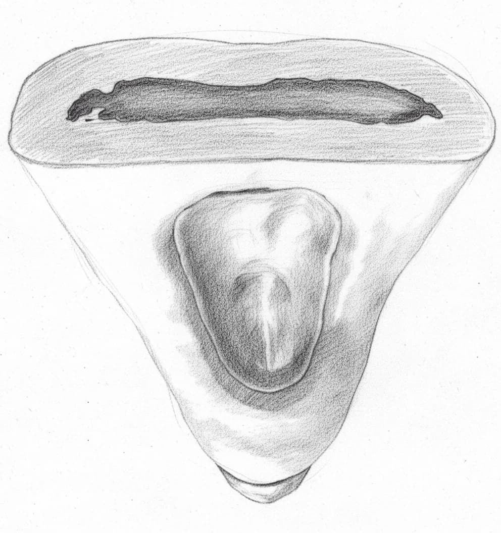
Mandibular Central Incisor - Axial View showing oval access cavity
Average Length20.7 mm
Root Development10 years
Universal #24, 25
FDI #31, 41
Access Cavity Design
ShapeTriangular/Oval
OrientationBuccolingual (NOT mesiodistal) — aligns with the oval canal cross-section; extend into cingulum
Entry PointLingual surface, just above cingulum
Landmarks
- Cingulum as inferior boundary — extend into it to find lingual canal
- Lingual shelf (dentinal bulge) must be removed
Do Not Invade
- Labial wall (thin dentin — maintain structural integrity)
- Incisal edge (preserve esthetics)
Canal Anatomy
Configurations
Single canal (Vertucci Type I)55-87%
Two canals merging (Vertucci Type II, 2-1)10-25%
Two canals with two foramina (Vertucci Type IV, 2-2)1-5%
Other (Type III, V)<5%
Canal Positions
BBuccal Canal
Easier to locate, more accessible; wider buccolingually than mesiodistally
LLingual Canal13-45% (ethnic-dependent)
Hidden beneath lingual shelf/cingulum — most commonly missed canal in mandibular anterior teeth
Danger Zones
- ⚠Lingual shelf — dentinal bulge hiding lingual canal orifice; must be removed for exploration
- ⚠Thin labial dentin — perforation risk if access directed too far labially
- ⚠Small tooth size — among the smallest teeth requiring endodontic treatment
Clinical Tips
⚠️
Warning
Extend access gingivally into cingulum to expose lingual canal — the lingual shelf hides it
🔧
Technique
Remove lingual shelf (dentinal bulge) sufficiently — it shields the lingual canal orifice from direct view
🔧
Technique
Use 20-30 degree distal angled radiograph to separate buccal and lingual canals
💡
Tip
If file appears off-center on straight PA, suspect second canal — confirmed clinical indicator
💡
Tip
Smallest tooth to treat endodontically — use small access burs, magnification highly recommended
Anatomical Variations
Vertucci Type II (2-1 configuration)
10-25%Two canals (buccal and lingual) originating separately but merging before the apex into a single foramen. Isthmus harbors tissue.
Access Modification: Extend access into cingulum; remove lingual shelf; thorough irrigation of isthmus region
Vertucci Type IV (2-2 configuration)
1-5%Two separate canals from chamber to apex, each with its own foramen. Requires independent treatment of each canal.
Access Modification: Extended buccolingual access to visualize both orifices; separate obturation for each canal