MandibularTrapezoidal
Mandibular First Molar
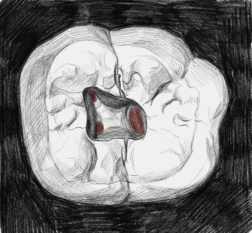
Mandibular First Molar - Axial View showing trapezoidal access with MB, ML, and distal canal orifices
Average Length21.0 mm
Root Development9-10 years
Universal #19, 30
FDI #36, 46
Access Cavity Design
ShapeTrapezoidal/Rectangular
OrientationBase faces mesial, shorter side faces distal
Entry PointMesial two-thirds of occlusal surface
Do Not Invade
- Mesial marginal ridge
Canal Anatomy
Configurations
2 roots, 3 canals (MB, ML, D)~67%
2 roots, 4 canals (MB, ML, DB, DL)~28-30%
3 roots (Radix Entomolaris)3-6% globally; 5-30% in East Asian populations
Canal Positions
MBMesiobuccal
Slightly buccal to mesiobuccal cusp tip
MLMesiolingual
Slightly lingual, connected to MB by isthmus
DDistal
Central when single (oval), or DB + DL when two
MMMiddle MesialUp to 46.2% with magnification
Between MB and ML, often below pulp floor level
RERadix Entomolaris3-6% globally; 5-30% in East Asian populations
Extra distolingual root with sharp buccal curvature
Danger Zones
- ⚠Mesial furcation perforation
- ⚠Stay within mesial two-thirds of occlusal surface
Clinical Tips
⚠️
Warning
Access shape is TRAPEZOIDAL/RECTANGULAR, NOT triangular — wider mesially
🔧
Technique
Middle mesial canal (MMC): anatomical prevalence 5-15%; detection up to 46.2% with microscope + troughing
💡
Tip
Microscope increases MMC detection from ~7% (naked eye) to ~46% — magnification transforms outcomes
🔧
Technique
For Radix Entomolaris: Extend access distolingually; flexible NiTi files essential for sharp buccal curvature
🔧
Technique
SLOB technique + 20-30 degree mesial or distal angulation reveals superimposed RE on PA
Anatomical Variations
Middle Mesial Canal (MMC)
Up to 46.2% with magnificationBetween MB and ML canals, often only accessible below pulp floor
Access Modification: Trough along developmental groove connecting mesial canals
Radix Entomolaris
3-6% globally; 5-30% in East AsianExtra distolingual root with sharp buccal curvature and apical 'kick'
Access Modification: Extend distolingually, trapezoidal shape with distal base
Ethnic Variation
East Asian (Chinese, Korean, Eskimo)5-30%
Caucasian, African, Indian<5%