MandibularTriangular
Mandibular Lateral Incisor
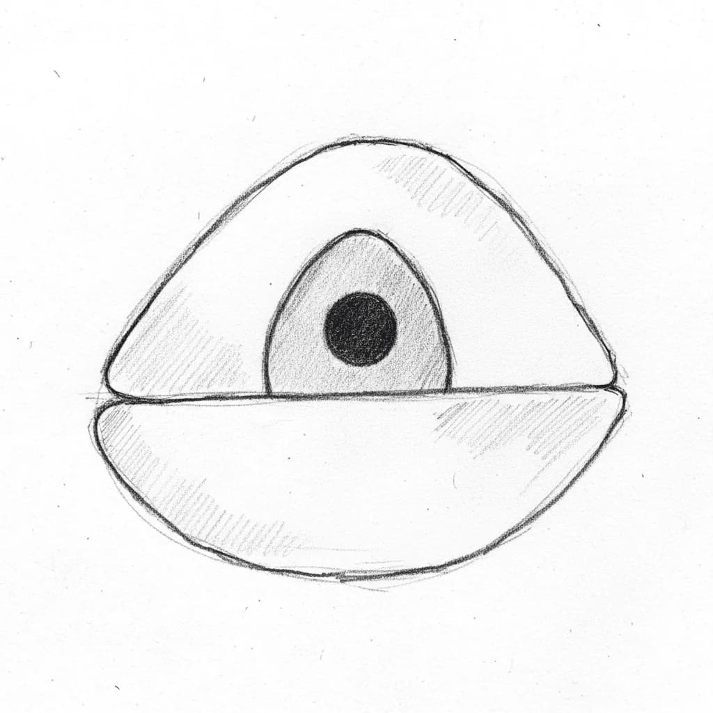
Mandibular Lateral Incisor - Axial View showing oval access cavity
Average Length21.1 mm
Root Development10 years
Universal #23, 26
FDI #32, 42
Access Cavity Design
ShapeTriangular/Oval
OrientationBuccolingual (NOT mesiodistal) — same as central incisor; extend into cingulum
Entry PointLingual surface, just above cingulum
Landmarks
- Cingulum as inferior boundary — extend into it
- Lingual shelf must be removed for exploration
Do Not Invade
- Labial wall (thin dentin — maintain structural integrity)
- Incisal edge (preserve esthetics)
Canal Anatomy
Configurations
Single canal (Vertucci Type I)60-75%
Two canals merging (Vertucci Type II, 2-1)15-25%
Two separate canals (Vertucci Type IV, 2-2)1-5%
Other (Type III, V)<5%
Canal Positions
BBuccal Canal
Easier to locate; wider buccolingually than mesiodistally
LLingual Canal25-40%
Hidden beneath lingual shelf — more commonly present than in central incisor
Danger Zones
- ⚠Lingual shelf — same anatomy as central incisor; must be removed for lingual canal exploration
- ⚠Thin labial dentin — perforation risk during access
- ⚠Root curvature more prevalent than central incisor (up to 70%) — instrument fatigue risk
Clinical Tips
🔧
Technique
SLOB Rule: Same Lingual, Opposite Buccal for canal identification on angled radiographs
🔧
Technique
Pre-curve small files (#10, #15) and explore along both buccal and lingual walls separately
🔧
Technique
Same lingual shelf anatomy as central — extend access into cingulum, remove shelf with ultrasonics
💡
Tip
Slightly longer than central (~21.1 mm vs ~20.7 mm) — adjust working length accordingly
Anatomical Variations
Vertucci Type II (2-1 configuration)
15-25%Two canals (buccal and lingual) merging before apex into single foramen. More common than in central incisor.
Access Modification: Extend access into cingulum; remove lingual shelf; irrigate isthmus thoroughly
Vertucci Type IV (2-2 configuration)
1-5%Two completely separate canals with independent foramina. Requires separate treatment of each canal.
Access Modification: Extended buccolingual access; separate obturation for each canal